Call Us Today!
(631) 821-2244

Diabetic eye disease describes a set of eye conditions that develop as a result of long-term diabetes and elevated blood sugar. These conditions — which commonly include diabetic retinopathy, diabetic macular edema, cataracts and glaucoma — can gradually impair vision or lead to sudden changes in sight if left unchecked. Understanding the connection between systemic metabolic health and eye structure is the first step toward protecting long-term vision.
Damage occurs because high glucose levels affect the tiny blood vessels and tissues inside the eye. In the retina, fragile vessels can leak or close off, depriving retinal tissue of oxygen and triggering harmful changes. The macula, the central part of the retina responsible for sharp vision, is especially vulnerable to swelling from fluid buildup, which can significantly reduce the clarity needed for reading, driving and recognizing faces.
Although these processes are common among people with both type 1 and type 2 diabetes, the pace and severity of eye involvement vary widely between individuals. Factors such as blood sugar control, blood pressure, duration of diabetes and overall vascular health influence risk. Because symptoms can be subtle in early stages, proactive monitoring is essential for preserving vision.
Annual dilated eye exams are the cornerstone of preventing preventable vision loss from diabetes. Dilation expands the pupil so eye care professionals can view the full retina and optic nerve clearly — areas where early signs of diabetic damage typically appear. Detecting abnormalities before they affect central vision makes successful treatment more likely and less invasive.
Beyond spotting blood vessel changes, dilated exams allow clinicians to identify early cataract formation and optic nerve changes consistent with glaucoma. These conditions are often manageable when found early, but they can progress silently. The exam also establishes a baseline record that helps your optometrist compare changes over time and tailor follow-up frequency to your individual risk.
For many patients, more frequent monitoring is recommended when retinopathy or macular swelling is present, or when systemic risk factors are out of range. Timely follow-up appointments and ongoing communication with your primary care provider or endocrinologist help ensure eye care keeps pace with your overall diabetes management.
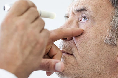
A diabetic eye exam is comprehensive and focused on detecting even subtle signs of disease. The visit typically begins with a review of medical history and medications, followed by testing of visual acuity and eye pressure. Pupil dilation is used so the doctor can examine the retina and macula in detail. These steps are painless and fundamental to a thorough assessment.
Modern diabetic eye exams often include advanced imaging alongside the clinical examination. Optical coherence tomography (OCT) produces high-resolution cross-sectional images of the retina, revealing macular swelling and thinning that aren’t always visible on routine inspection. Wide-field retinal photography can document areas of the retina affected by bleeding or vessel closure, creating a permanent record for monitoring progression.
Based on findings, the doctor will discuss the results and recommend a personalized follow-up plan. That may mean routine observation, sooner rechecks, or referral to a retinal specialist for further evaluation. Patient education is a key component of the visit — understanding test results and next steps empowers patients to participate in protecting their vision.
Eye exams are critical, but preventing progression of diabetic eye disease requires attention to daily health habits and coordination with your broader medical team. Tight control of blood glucose is one of the most important modifiable factors. Consistently maintaining recommended blood sugar targets reduces stress on the delicate vessels inside the eye and lowers the chance of disease progression.
Blood pressure and cholesterol also play powerful roles in determining eye outcomes. Hypertension and high lipid levels accelerate vascular damage and can worsen retinal disease. Regular monitoring and treatment of these conditions, combined with a balanced diet, physical activity, and smoking cessation when applicable, form a practical plan for reducing ocular risk.
Equally important is knowing when to seek care. Any sudden change in vision — new floaters, flashes of light, dark patches, or rapid blurring — should prompt an immediate evaluation. Even subtle fluctuations deserve attention; early intervention can preserve function and expand treatment options.
Treatment for diabetic eye disease is tailored to the specific condition and severity. For diabetic macular edema and certain stages of retinopathy, intravitreal medicines that reduce swelling and inhibit abnormal vessel growth have become standard. In other situations, laser therapy can seal leaking vessels or reduce the risk of bleeding in the retina. Cataracts and glaucoma are managed with established surgical or medical therapies when they affect vision or threaten eye health.
Successful treatment often depends on timely identification and adherence to follow-up plans. Many therapies require a series of visits or repeated treatments over months to achieve the best result. Your eye care team will monitor response to therapy with clinical exams and imaging, adjusting the plan if needed to optimize visual outcomes.
Coordination between eye care providers and your primary care or diabetes specialists is essential after a diagnosis. Adjustments to systemic medications, tighter control of cardiovascular risk factors, and close monitoring can all influence the course of eye disease. Working together as a team gives patients the best chance to maintain useful vision for years to come.
At Soundview Eye Center we take diabetic eye disease seriously and focus on early detection, clear communication and individualized care plans. Our goal is to help patients understand their condition and take informed steps to protect their vision while coordinating with their broader health team. Contact us for more information or to discuss how we can support your eye health and diabetes management.
Diabetic eye disease describes a group of eye conditions that develop as a result of long-term diabetes and elevated blood sugar. Common examples include diabetic retinopathy, diabetic macular edema, cataract and glaucoma. These disorders can slowly reduce vision or cause sudden changes if not detected and managed early.
High glucose damages the tiny blood vessels and supporting tissues inside the eye, causing leakage, vessel closure and abnormal vessel growth. When the macula swells with fluid, central vision used for reading and recognizing faces can become blurred or distorted. Because early changes are often symptomless, regular monitoring is essential to protect long-term vision.
People with both type 1 and type 2 diabetes should have regular dilated eye exams to screen for complications. For many adults with diabetes, an annual dilated exam is a common recommendation, but schedules are individualized based on disease severity, control and other risk factors. Newer onset or well-controlled disease may require different timing than long-standing or unstable diabetes.
Patients who have any degree of retinopathy, macular swelling, poor systemic control or other ocular findings typically need more frequent follow-up. Pregnant patients with diabetes often require earlier and closer monitoring because pregnancy can accelerate retinal changes. Your eye doctor will recommend a personalized interval based on exam findings and overall health.
A diabetic eye exam begins with a review of medical history and medications followed by measurement of visual acuity and intraocular pressure. Pupillary dilation allows the doctor to examine the retina and optic nerve thoroughly, which is critical for detecting subtle signs of disease. These steps are painless, though dilation can temporarily blur near vision and increase light sensitivity.
Most visits include discussion of symptoms, risk factors and a clear plan for follow-up based on findings. Advanced imaging such as optical coherence tomography and retinal photography may be performed to document and quantify changes. At Soundview Eye Center our team reviews results with patients and explains next steps so they understand their condition and treatment options.
Optical coherence tomography (OCT) produces high-resolution cross-sectional images of the retina and is especially useful for detecting and measuring macular swelling. Wide-field retinal photography and fundus imaging create a permanent record of the retinal surface, helping clinicians spot areas of bleeding or vessel closure that might be missed on a basic exam. In selected cases, fluorescein angiography is used to map circulation and identify leaking or non-perfused areas.
Standard clinical testing also includes visual acuity, slit-lamp examination and intraocular pressure checks to screen for cataract and glaucoma. Together, clinical exam and imaging form a comprehensive assessment that guides treatment decisions and monitoring frequency. Imaging makes it easier to track subtle changes over time and to communicate findings clearly with other members of a patient’s care team.
Chronic high blood sugar directly harms the microvasculature of the retina, increasing the risk of leakage, ischemia and abnormal vessel growth. Maintaining glucose levels within recommended targets reduces stress on retinal vessels and lowers the likelihood of progression. Good systemic control does not eliminate all risk, but it is one of the most powerful modifiable factors for protecting vision.
Elevated blood pressure and abnormal cholesterol levels accelerate vascular damage and worsen retinal disease by promoting vessel leakage and occlusion. Coordinated management with your primary care physician or endocrinologist to control blood pressure, lipids and glucose is a key component of an eye-protection strategy. Lifestyle measures such as a balanced diet, regular activity and smoking cessation also support ocular health.
In many cases early diabetic eye disease causes no noticeable symptoms, which is why routine dilated exams are so important. When symptoms do occur they can include blurring of central vision, sudden floaters or flashes of light, areas of shadow or a general decline in contrast and color perception. Any new or rapidly worsening visual changes should prompt timely evaluation.
Because gradual changes may be subtle, patients are encouraged to report even small fluctuations in vision rather than waiting for persistent loss. Prompt detection expands treatment options and improves the chance of preserving useful vision. Keep a record of symptom onset and describe it during your eye visit so clinicians can correlate symptoms with exam findings.
Treatment depends on the type and severity of disease; for diabetic macular edema and many forms of sight‑threatening retinopathy, intravitreal medications that reduce swelling and inhibit abnormal vessel growth are commonly used. Laser photocoagulation remains an important option to seal leaking vessels or reduce the risk of bleeding in specific circumstances. These treatments are chosen based on exam findings and imaging results.
Advanced or non‑resolving cases may require surgical intervention such as vitrectomy, and coexisting cataract or glaucoma are managed with established surgical or medical therapies when they threaten vision. Many therapies require a series of visits and close monitoring to assess response and adjust the plan. Your eye care team will explain the benefits, typical course and expected follow-up so you know what to expect.
Because diabetic eye disease is linked to overall vascular health, collaboration between eye care providers and systemic care teams improves outcomes. Sharing information about glycemic control, blood pressure, lipid levels and medication changes allows eye doctors to interpret retinal findings in the context of the patient’s broader health. Timely communication helps align treatment priorities and follow-up schedules.
When retinal disease is detected, coordinated care may include medication adjustments, expedited referrals and closer monitoring during periods of systemic instability such as pregnancy or major medication changes. Patients benefit when all providers work together to reduce risk factors and support adherence to recommended monitoring and therapy. Ask your eye doctor if they will send exam summaries to your primary care provider or endocrinologist to keep everyone informed.
Bring a current list of medications, any recent lab results such as a recent HbA1c if available, and a brief medical history that highlights changes since your last visit. Arrive with any glasses you use for distance and reading so the clinician can document your baseline function. Sharing symptoms, vision changes and dates of any other eye treatments helps the team build a complete picture.
Because dilation may blur near vision for several hours, consider arranging transportation or planning activities accordingly. There is no special fasting required for the eye exam itself, but be ready to discuss your diabetes control and any recent systemic changes. Being prepared helps the visit run smoothly and supports accurate diagnosis and follow-up planning.
Soundview Eye Center combines experienced clinicians, advanced retinal imaging and a patient-centered approach to detect and manage diabetic eye disease early. Our providers focus on clear communication, individualized care plans and coordination with each patient’s medical team to ensure ocular findings are interpreted within the larger context of diabetes management. That integrated approach helps tailor monitoring and treatment to each patient’s needs.
We strive to make exams informative and actionable by explaining results, imaging and next steps in plain language so patients can participate in decisions about their care. Patients in the Shoreham, NY area can expect thorough dilated examinations, appropriate imaging and a plan for follow-up that reflects current clinical best practices. If you have questions about scheduling or what to bring to your appointment, your eye care team can provide guidance.